AG Lochnit - other model systems
Ascaris suum, an intestinal parasite
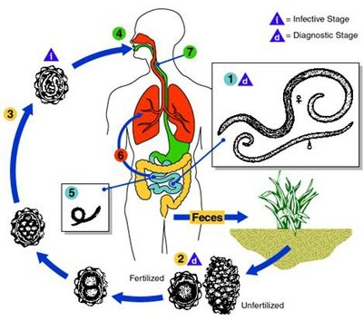
There has been much debate on the taxonomic status of human and pig Ascaris. Scanning electron microscopy and biochemical differences have finally indicated two separate species, A. lumbricoides and A. suum, respectively. Due to their close relationship, they are regarded as sibling species (1). The parasite shows a simple life cycle (2). Eggs are excreted in the feces, with the infective larvae developing within 9-13 days. After the eggs are swallowed, the larvae hatch in the duodenum. The L2 larvae burrow into the mucosa, penetrate blood vessels and appear in the liver after 6 hours. In the liver they develop to the L3 stage within a few days. They then migrate to the heart and lungs. After seven days they break into the alveoli and reach the intestine via the trachea, where they molt again, reach adulthood, mate and produce eggs.
Ascaris has been used as the prototypic nematode model organism for decades, due to its large size (females 200-350 nm, males 150-310 mm), in biochemical and neurophysiological studies. Since large quantities of worms can be obtained at the abattoir, isolation of biomolecules is easily feasible. In vitro cultivation of adult nematodes is possible and also generation of early adults from eggs in vitro has been reported (3-5). Due to its large size and well known anatomy, this nematode is an excellent model for immunofluorescence and immunohistochemical studies.

copyright: private
Acanthocheilonema viteae, a model for filariasis
Due to the problems of cultivating human filariids like Onchocerca volvulus or Wuchereria bancrofti in animal models, A. viteae, a rodent filariid, has become one of the most studied model systems for filariasis (6,7). A. viteae contains PC-substituted antigens similar to those of the human filarids (8-10). Adults recovered from jirds can be maintained for days in cell culture medium, thus allowing metabolic studies. The jird, Meriones libycus, is usually infected by subcutaneous injection of L3 larvae obtained from the tick Ornithodorus moubata. Microfilariae appear in the blood circulation after 42-65 days after infection. Ticks were infected by allowing them to feed on the shaved abdomen of anesthetized jirds. After two molts they develop within 6 weeks to the infective stage.
Literature:
1. Nadler, S. A. (1987) Journal of Parasitology 73, 811-816
2. Crompton, D. W. T. (1989) in Ascaris and its prevention and control (Crompton, D. W. T., Nesheim, M. C., and Pawlowski, Z. S., eds), pp. 9-44, Taylor & Francis, London
3. Levine, H. S., and Silverman, P. H. (1969) J. Parasitol. 55, 17-21
4. Douvres, F. W., and Urban, J. F. (1983) J. Parasitol. 69, 549-558
5. Douvres, F. W., and Urban, J. F. (1986) Proc. Helminthol. Soc. Wash. 53, 256-262
6. Worms, M. J., Terry, R. J., and Terry, A. (1961) Journal of Parasitology 47, 963-970
7. Eisenbeiss, W. F., Apfel, H., and Meyer, T. F. (1991) J Parasitol 77, 580-586
8. Gualzata, M., Weiss, N., and Heusser, C. H. (1986) Exp. Parasitol. 61, 95-102
9. Gualzata, M. D., Rudin, W., Weiss, N., and Heusser, C. H. (1988) Parasite Immunol. 10, 481-491
10. Harnett, W., Worms, M. J., Kapil, A., Grainger, M., and Parkhouse, R. M. E. (1989) Parasitology 99, 229-239
