Gallery of pictures from 2011
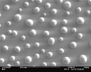
The SEM picture shows an originally dense and covering thin silver layer, which was deposited by pulsed laser deposition (PLD) on top of an (111) oriented substrate of yttria stabilised zirconia (YSZ). In the picture the dewetted surface after annealing the sample at 400 °C for 1 hour in oxygen atmosphere is shown. Although silver has a melting point of 962°C, thin silver films are unstable even at moderate temperature, as silver shows an extremely high mobility. Picture submitted by C. Raiß
Interior view of the new MACCOR multipotentiostat: Providing 96 independent channels, the system allows to execute automatically computer-controlled electrochemical experiments to measure the performance of a large variety of different battery cells actually investigated in the group of Prof. Janek. The increase to 96 independent channels allows to get time-efficiently an insight into the cell performance and its dependencies to the cycling parameters. The MACCOR system represents an important supplementation to the present laboratory equipment. Picture submitted by Martin Busche
Cross-section of bone: A TOF-SIMS Image recorded within the SFB-Transregio Project 79. The Justus-Liebig-University, the Technical University of Dresden and the University of Heidelberg are working together to develop and test new material concepts for tissue regeneration in systematically diseased bones. The AG-Janek participates in this project i.a. with 3-D analytics of the implant interface in osteoporotic bones. The picture shows a TOF-SIMS image from an implantation cross section investigated within the framework of the SFB-Transregio-project 79. The cross section has been provided and prepared by the department of trauma surgery of the university hospital Giessen Marburg. The TOF-SIMS image shows the intensity distribution of the sum of the PO2-, PO3-, C16H31O- and C16H29O- signals on an area of 500 µm times 500 µm. The signal is normed with the total signal intensity distribution.The SIMS image shows besides the implant (1) typical components of bone tissue such as osteoid (2), osteoblasts (3), erythrocytes (4) and adipocellulars of bone marrow (5). Picture submitted by A. Henß
Rediscovery of Alchemy. Production of gold sucessful accomplished. In the night of the 1st April it was possible after long lasting experiments to produce gold from silver. A silver ion conductor (silver iodide, AgI) was used to create with a high voltage field high reactive silver atoms on the surface of the ion conductor. In addition a great flux of neutrons was directed to the surface area. The silver cores are excited due to the neutron bombardement and the cores reorganice to gold. In the end a gold film on the surface is created with small impurities of aluminium, silicium or phosphor. Picture for April Fool's joke.
Since beginning of February the Institute of Physical Chemistry possesses a new high resolution scanning electron microscope (HRSEM) called MERLIN from ZEISS. It has a nominal resolution of 0.8 nm and is equipped with six detectors: Two SE detectors for raster images of sample surfaces; two BSE detectors for material contrast investigations; an EDX detector from OXFORD INSTRUMENTS for sample composition analysis; and an EBSD system for crystal orientation analysis. Furthermore, it is equipped with a charge compensation for insulating samples. Picture submitted by: Dr. Klaus Peppler (klaus.peppler @ phys.chemie.uni-giessen.de).
A new machine for pulsed laser deposition (PLD) of thin films arrived at the Institute of Physical Chemistry in the beginning of February. It is equipped with two lasers with different wavelengths: an excimer laser in the UV range and a Neodym-YAG laser in the infrared range. In addition to the different wavelengths for ablation the machine is equipped with two systems for substrate heating: a resistive heater and an infrared laser heater for direct heating of the substrate rear side. The targets with primary material are assembled in a rotating table with 5 positions. These targets are positioned into the laser beam automatically according to a previously programmed process. Likewise the background pressure is adjusted automatically by mass flow controllers. A manipulator bar allows direct gating out of and into a glove box attached to the machine. Thus handling of air sensitive thin films is possible. Picture submitted by A. Braun
The Federal Ministry of Education and Research invests 5.5 million euro into the project “IN-TEG”. Material scientists at the Justus-Liebig-University of Giessen receive about 800.000 euro. The research group of Prof. Janek is a part of the project IN-TEG with seven other partners from science and industry. IN-TEG is a research consortium which investigates new thermoelectric materials for energy transformation. The picture shows some of the project partners during the presentation of the funding decision. The funding documents were handed over by Dr. Helge Braun (secretary of state, first left). Next to Prof. Janek (second left) on the right stands Dr. Kerstin Schierle-Arndt (BASF), Dr. Eckhard Müller (DLR), Prof. Barbara Albert (TU Darmstadt) and Prof. Juri Grin (MPI Dresden). The picture was kindly provided by the press office of the JLU. The whole press release in German can be found here: http://www.uni-giessen.de/cms/ueber-uns/pressestelle/pm/pm162-11/
Hunting of rotten bones – The Janek group is a member of the DFG Sonderforschungsbereich TRR79, which has the aim to develop new implant materials for osteoporosis patients. The shown image is from an article of the journal labor&more (August 2011). From the left to the right: Prof. Dr. Christian Heiß, Prof. Dr. Jürgen Janek, Dr. Anja Henß, Dr. Marcus Rohnke
Since May, a spectrometer of the type PHI 5000 VersaProbe™ is available for Electron Spectroscopy for Chemical Analysis (ESCA) at the Institute of Physical Chemistry. This facility offers analysis methods like X-Ray photoelectron spectroscopy (XPS), Ultra-violet photoelectron spectroscopy (UPS), and Auger electron spectroscopy (AES). By sputtering with an Argon or C60 source, it is possible to measure depth profiles of a sample. An aluminium anode is used to create a monochromatic X-ray beam for XPS measurements. This beam can be focused (<10 μm) on points of interest of the sample surface also enabling to create lateral maps of the sample composition. The region to be analyzed can be chosen by previously recording scanning X-ray (SXI) or scanning electron microscopy (SEM) images. Furthermore, it is possible to perform angle-resolved measurements (AR-XPS). Picture submitted by H. Metelmann
The picture shows Dipl.-Chem. Manuel Pölleth after completing his PhD oral exam. In his dissertation Manuel Pölleth has studied electrochemical processes at the interface between a plasma and a liquid. In this process he has deposited silver nanoparticles at this interface to investigate the influence of various physical properties on the particle size distribution and morphology, e.g. viscosity and surface tension of the liquid,. Due to their low vapor pressures ionic liquids were particularly suitable as a template for such a plasma-electrochemical deposition. Picture submitted by M. Göttlicher
Sodium-lab opened: A new laboratory dedicated to the research on sodium electrochemistry was opened. The picture shows the recently acquired glovebox (4 working places) in which air sensitive experiments such as the assembly of sodium cells will be conducted. The lab was also equipped with an elemental analysis machine and a tubular oven for heat treatments of samples under defined atmospheres. Along with the research on sodium batteries, the lab will be also used to prepare engineered carbon materials. An article on this topic was recently published in "Energy & Environmental Science".
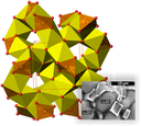
The figure shows a part of the crystal structure of Li7La3Zr2O12 garnet (LLZO), showing the 3D connected channels responsible for Li+-transport in LLZO. The 3D-connected space consists of interstitial octahedral sites (yellow) sharing two opposite faces with neighbouring “LiO4” tetrahedral sites (orange). The inset shows LLZO grains with a novel Li+-conducting phase in the grain boundaries. The interfacial Li+-conducting phase enhances the Li+-conductivity of LLZO, reaching a value of 5.4 ⋅ 10−4 S⋅cm−1 at 20 °C. Since August 2011 Dr. Hany El-Shinawi works as Fellow of the Alexander-von-Humboldt Foundation at the Institute of Physical Chemistry in the group of Prof. Janek. Dr. El-Shinawi studied chemistry at the Mansoura University in Egypt and obtained his PhD at the University of Birmingham. He is a specialist in the preparation and characterization of functional oxides and currently studies a number of solid lithium-ion conductors. As a member of the solid-electrolyte team in Prof. Janek group, Dr. El Shinawi is particularly trying to develop and characterize novel garnet-type lithium electrolytes. The study also aims to optimize the structural and electrochemical properties of these garnets for their use as electrolytes in all-solid-state batteries. For further information about our latest activities in this field we refer to the publication “Structure and dynamics of the fast lithium ion conductor ‘Li7La3Zr2O12’” by H. Buschmann, J. Dölle, S. Berendts et al., Phys. Chem. Chem. Phys. 13 (2011) 19378-19392.


