AG Lochnit - C. elegans
In 1965, Sidney Brenner selected C. elegans as a model system to study animal development and behavior [1]. The short life cycle, small size, ease of laboratory cultivation and genetic manipulation has made this worm to the best understood metazoic organism today. The C. elegans population consists mainly of hermaphrodites and only a small number of males. The life cycle consists of 14 hours embryogenesis and 36 hours of postembryonic development through four larval stages. The hermaphrodite produces sperm at the late L4 stage, then returning to the production of oocytes as an adult. Progeny are restricted to 300-350 individuals due to the limited number of sperm, but can be raised to 1000 by mating with males. Under unfavourable food conditions the nematode can undergo dauer larva formation at the second molt. Dauer larvae can survive for months. The hermaphrodite consists of 959 cells with 302 nerve cells deriving from four somatic and one germ-line cell. The complete wild-type cell lineage from the fertilized egg to the adult was determined by observation of cell division and cell migrations in living animals due to the transparency of the body [2]. The genome was completely sequenced in 1998, indicating approximately 20,000 genes [3]. The large number of laboratories working on C. elegans has led to the establishment of several platforms providing the scientific community with information and material. The WormBase contains information on genomic sequences, mutants, phenotypes, strains, RNA interferance (RNAi) experiments and gene homologies. The ORFeome project provides all open reading frames in referential vectors allowing convenient transfer in different expression systems. A DNA microarray service is offered by the Sanger Institute, Cambridge England and a complete E. coli RNAi feeding library covering all ORFs by the MRC. Since this nematode shows high conservation in anatomy, genetics and development, C. elegans can be regarded as an excellent model even for parasitic species [4-7].

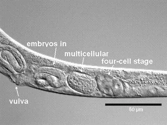
Anatomy of C. elegans copyright: private

Anatomy of C. elegans 2 copyright: private
Literatur:
[1] Brenner S. The genetics of Caenorhabditis elegans. Genetics 1974;77:71-94.
[2] Sulston J, Horvitz HR, Kimble J. Appendix 3. Cell lineage. In: Wood, W.B. and Reasearchers, T.C.o.C.e., The nematode Caenorhabidits elegans. Cold Spring Harbour Laboratory, Cold Spring Harbour, New York: 1988:457-89.
[3] consortium TCes. Genome sequence of the nematode C. elegans: a platform for investigating biology. Science 1998;282:2012-8.
[4] Bird AF, Bird J. (1991) The structure of nematodes. Academic Press, San Diego.
[5] Szymanski M, Opiola T, Barciszewska MZ, Barciszewska J. The nucleotide sequence of 5S ribosomal RNA from the parasitic nematode Ascaris suum. Evolutionary relationships in nematodes. Biochim Biophys Acta 1996;1308:251-5.
[6] LaVolpe A. A repetitive DNA family, conserved throughout the evolution of free-living nematodes. J Mol Evol 1994;39:473-7.
[7] Sommer RJ, Carta LK, W. SP. The evolution of cell lineage in nematodes. Development 1994;Suppl.:85-95.
